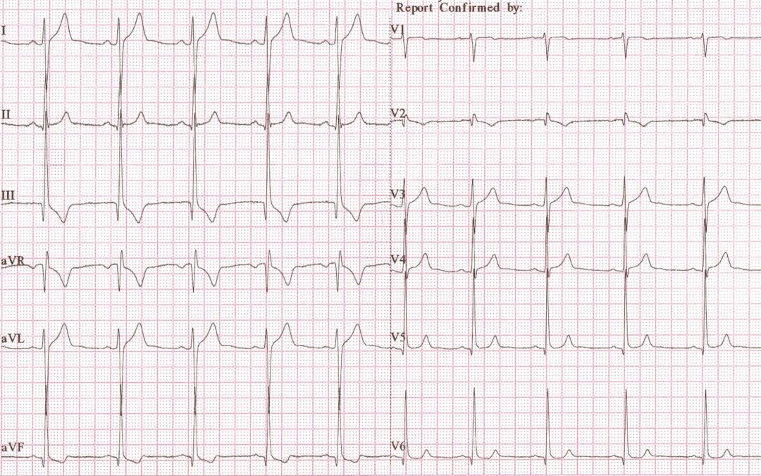
This ECG was recorded from a 21-year old female who had complained of palpitations. On examination she had an ejection systolic murmur.
What is the most likely explanation for the abnormal ECG appearances?
Answer:
Well, it looks like my clue was useful to a few people. Others may have recognised the pattern or worked it out for themselves, but what happened is that the left arm and V2 connections were swapped for this ECG. The typical features of this technical error have been documented but I suspect that it usually goes unrecognised. The key ECG characteristics, all on show in this recording, are:
- Deep S waves in leads I and aVL
- Tall R waves in leads III and aVF
- T wave inversion in III and aVF
- Lead V2 looks ‘wrong’ and doesn’t fit in with the usual progression from V1 to V3. In our example, V2 has a negative T wave whereas its immediate neighbours, V1 and V3, both have a positive T wave.
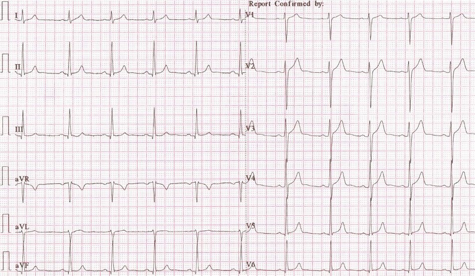
Note that lead II is entirely unaffected by the error; the other 11 leads are all altered. The effects of this connection swap can easily be appreciated when you compare the original ECG with this one, which was recorded after the error had been corrected. It is now completely normal.
Dr Dave Richley,
Associate Lecturer
Newcastle University
Hi Dave – I was just about to launch into an analysis/possible causes of LVH with a right axis deviation but there is just something odd about this ECG I can’t quite put my finger on. Maybe it’s the format I’m not used to looking at. Just checking but are the leads on correctly? The P wave in lead I appears slightly larger than the P wave in lead II so I’m wondering if this is a left arm/left leg swap? Lead III just doesn’t look quite right to me, and V2 also looks a bit strange. If leads are on correctly I’ll post what I originally thought. Many thanks.
Hi Max – You’re right to be suspicious about the electrode connections, but what exactly has been done?
Her’s a clue: the title of a song by Coldplay.
I have looked at this ECG many times since it was posted & I also thought it looked strange. I would have probably said something like LVH with strain pattern but V2 completely through me.
With your Coldplay hint ? Yellow. I’ll take a stab at, the left arm lead being placed in the V2 position? As they both can be seen as the ‘yellow’ lead on some machines?
I’m glad someone else thought the ECG didn’t look right! Trying to work it out as the patterns just seem all wrong.
The murmur is suggestive of outflow tract obstruction – HCM, aortic or possibly pulmonary stenosis
Left arm – V2 electrode swap?
V2 and LA swap? A muddling of the yellows I’m guessing from the clue!
Left arm switched with left leg?